When you notice something unusual growing from an older family member’s skin, it can be difficult not to panic.
That is especially true when the growth looks almost nothing like an ordinary mole, pimple, or skin tag.
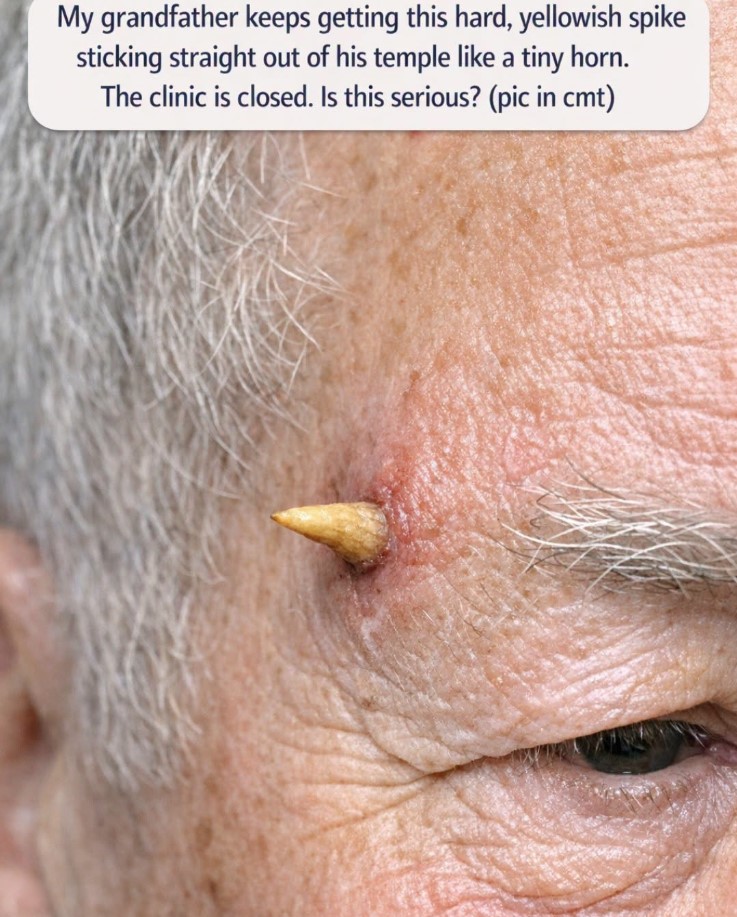
In this case, the concern was a hard, yellowish projection sticking straight out from an older man’s temple. It looked almost like a miniature horn growing from the skin.
The first instinct might be to wonder whether it is simply a strange wart or a piece of hardened skin.
And when the clinic is closed, the next temptation may be even stronger: remove it yourself and deal with the problem before morning.
But this is one situation where trying to cut, pull, scrape, or burn the growth away at home is not a good idea.
A horn-like growth on the skin can be something called a cutaneous horn. The visible projection itself is made primarily of compacted keratin, the same tough protein found in the outer layer of skin and in fingernails.
The important part, however, isn’t necessarily the horn that you can see.
It is the skin underneath it.
That base can sometimes be associated with a harmless skin growth, but it can also develop from a precancerous lesion or skin cancer. That is why a doctor needs to examine it properly rather than judging the growth only by its appearance.
What exactly is a cutaneous horn?
A cutaneous horn is a firm projection of keratin that rises above the surface of the skin.
It can look surprisingly similar to a tiny animal horn.
The color may range from:
- Yellow
- Cream
- White
- Tan
- Gray
- Brownish yellow
Some are very small, while others become considerably longer and more noticeable.
They can develop on different parts of the body, particularly areas that receive substantial sun exposure. The face, ears, scalp, backs of the hands, and forearms are among the locations where they can occur.
An older person’s temple is therefore a location worth taking seriously, particularly when there has been significant lifetime sun exposure.
But seeing a horn-like growth does not automatically mean someone has skin cancer.
That distinction is extremely important.
A cutaneous horn is a physical appearance, not a diagnosis by itself.
The underlying lesion is what determines whether the situation is harmless, precancerous, or malignant.
Why the base matters more than the horn
Imagine the visible horn as the tip of an iceberg.
The part you can see may simply be a thick buildup of keratin.
But underneath that keratin is the skin lesion that produced it.
That lesion can have several different causes.
Some cutaneous horns develop from benign conditions such as warts or seborrheic keratoses.
Others can be associated with actinic keratosis, a precancerous change related to cumulative sun damage.
Some can develop over squamous cell carcinoma, a type of skin cancer.
That is why doctors don’t simply look at the horn and decide, “It’s just dead skin.”
The underlying tissue needs to be evaluated. In some situations, this means removing the lesion or taking a biopsy so the tissue can be examined under a microscope.
Why the temple deserves attention
The temple is a part of the face that receives sun exposure throughout a person’s life.
For an older adult, that means decades of cumulative ultraviolet exposure may have affected the skin.
Sun damage can produce many different changes, most of which are not immediately dangerous. However, long-term sun exposure is also an important risk factor for several types of skin cancer and precancerous lesions.
So when an older person develops a new, hard, rapidly changing, horn-like growth on a sun-exposed area of the face, it is sensible to have it examined.
That doesn’t mean you should assume the worst.
It means you shouldn’t assume the best either.
The safest approach is to let a medical professional determine what is underneath the growth.
Is it an emergency tonight?
Usually, a stable cutaneous-horn-like growth without severe symptoms does not mean you need to perform an emergency procedure at home.
If your grandfather is otherwise feeling well, the growth isn’t heavily bleeding, and there is no severe pain, major swelling, fever, or other concerning symptom, the priority is generally to protect the area and arrange a medical evaluation as soon as reasonably possible.
A clinic being closed for one night does not mean you need to remove the growth yourself.
In fact, attempting to remove it could create a bigger problem.
The important thing is to recognize symptoms that would change the situation.
When should you seek urgent medical attention?
Pay attention to the condition of the skin around the growth.
Seek more urgent medical advice if there is:
- Bleeding that does not stop with steady pressure
- Rapidly increasing swelling
- Significant redness spreading around the area
- Pus or other concerning drainage
- Severe or rapidly increasing pain
- Fever or signs of infection
- A sudden major change in the lesion
- A problem involving the nearby eye or vision
These symptoms don’t automatically mean the lesion is cancerous.
They simply mean that waiting for a routine appointment may not be appropriate.
If there is significant bleeding, applying gentle, continuous pressure with clean gauze or a clean cloth can help while seeking medical advice.
Whatever you do, don’t cut it off
This is probably the most important advice for tonight.
If the growth looks like a little piece of horn or nail, it can be incredibly tempting to reach for nail clippers, scissors, tweezers, or a razor.
Don’t.
Cutting it off at home can cause bleeding, pain, and injury to the surrounding skin.
It also doesn’t solve the underlying problem.
Remember, the visible projection may only be the keratin portion.
The tissue at the base is what needs to be evaluated.
If someone simply cuts off the protruding portion, the underlying lesion remains.
There is also a risk that the procedure will make the area inflamed or irritated, making it harder for the clinician to assess what was originally there.
Don’t pull, twist, or sand it down either
Cutting isn’t the only problem.
Some people might think they can slowly twist the growth until it comes loose.
Others may consider filing it down with a nail file or using a pumice stone.
Those approaches aren’t appropriate either.
The growth isn’t something that should be treated like a thick toenail.
The goal isn’t to make the horn disappear temporarily.
The goal is to determine why it formed in the first place.
A medical professional can decide whether the entire lesion needs to be removed and whether the tissue should be sent for pathology.
Avoid wart removers and strong acids
It is also better not to experiment with over-the-counter wart treatments or strong chemical products on a facial growth.
Products containing acids can damage healthy skin around the lesion.
The skin of an older adult can also be thinner and more fragile than younger skin.
You don’t want to create a chemical burn on the temple while trying to treat something that hasn’t even been diagnosed.
Avoid experimenting with:
- Wart remover
- Salicylic acid products
- Freeze sprays
- Strong exfoliating acids
- Vinegar
- Essential oils
- “Drawing” salves
- Harsh disinfectants
None of these can tell you what is underneath the horn.
What can you safely do while waiting?
If the clinic is closed and the growth is stable, the safest approach is surprisingly simple.
Leave it alone.
Don’t pick at it.
Don’t squeeze it.
Don’t attempt to break it.
Don’t try to flatten it.
If it catches on clothing, bedding, glasses, or hair, you can protect the area gently with a small nonstick dressing.
The dressing shouldn’t press tightly against the growth.
Its purpose is simply to reduce the chance of accidentally snagging it.
This is particularly helpful overnight.
Someone can easily roll onto the affected side while sleeping and accidentally catch the growth on a pillowcase.
Keep the area clean
If the surrounding skin is dirty, gently clean the area with mild soap and water.
Don’t scrub the growth aggressively.
Pat the area dry rather than rubbing it with a towel.
If the skin isn’t broken, there is generally no reason to coat the lesion with multiple products.
Keeping things simple makes it easier to monitor any changes before the medical appointment.
What if it gets bumped and starts bleeding?
A horn-like growth can bleed if it is knocked or pulled.
If that happens, don’t panic.
Use clean gauze or a clean cloth and apply gentle, steady pressure to the bleeding area.
Don’t keep lifting the cloth every few seconds to see whether the bleeding has stopped.
Continuous pressure is more useful.
If bleeding continues despite sustained pressure or appears excessive, seek urgent medical advice.
This is particularly important if your grandfather takes medication that affects blood clotting, such as certain anticoagulants or antiplatelet medications.
In that situation, even a relatively small skin injury can sometimes bleed more than expected.
What does the doctor actually look for?
A dermatologist or other qualified clinician will examine the growth closely.
They may consider:
- How long it has been there
- How quickly it appeared
- How quickly it has grown
- Its size
- Its shape
- Its color
- The width of the base
- Whether the base is red or inflamed
- Whether it is tender
- Whether it bleeds
- Whether the surrounding skin has changed
The base is particularly important.
A horn that is relatively broad at its base, painful, inflamed, or growing quickly may warrant especially careful evaluation.
However, appearance alone cannot reliably tell you whether a lesion is benign or malignant.
Why a biopsy may be necessary
A biopsy allows a pathologist to examine the tissue under a microscope.
The doctor may remove the lesion completely or take an appropriate sample depending on its size, location, and appearance.
The important point is that the sample should allow evaluation of the base, not merely the hard keratin sticking outward.
That is because the keratin projection doesn’t tell the entire story.
The underlying skin is what can reveal whether the lesion is associated with a benign growth, actinic keratosis, squamous cell carcinoma, or another condition.
Could it simply be a wart?
Yes, a wart is one of the benign possibilities.
Some warts can become thick and keratinized and may look unusual.
But you shouldn’t assume that is what your grandfather has simply because the growth is hard.
The same general appearance can occur with several different underlying conditions.
That’s why an older person’s new horn-like lesion is better evaluated than treated blindly at home.
Could it be an age-related skin growth?
Another possibility is a benign growth such as a seborrheic keratosis.
These are extremely common in older adults and can have rough, thick, wart-like surfaces.
Some benign lesions can become heavily keratinized and contribute to a horn-like appearance.
Again, the appearance isn’t enough to make the diagnosis.
A dermatologist can often narrow down the possibilities by examining the lesion, but sometimes pathology is needed for certainty.
Could it be precancerous?
Yes.
One possible underlying cause of a cutaneous horn is actinic keratosis.
Actinic keratoses are associated with long-term ultraviolet exposure and often appear as rough, scaly, or thickened areas on sun-exposed skin.
Some can develop into squamous cell carcinoma, which is why they are often monitored or treated.
A horn-like projection developing from sun-damaged skin therefore deserves professional assessment.
Could it be skin cancer?
It is possible, but the presence of a cutaneous horn does not automatically mean cancer.
Some cutaneous horns arise from malignant or potentially malignant lesions, including squamous cell carcinoma.
Squamous cell carcinoma is particularly relevant because it commonly develops on chronically sun-exposed skin and can occur in older adults.
That is precisely why doctors don’t recommend simply clipping the horn off and forgetting about it.
The visible horn needs to be evaluated in context.
What makes a lesion more concerning?
There are several features doctors may pay particular attention to.
A lesion may deserve especially prompt evaluation if it:
- Has appeared recently
- Is growing quickly
- Has become painful
- Bleeds easily
- Has a thick or broad base
- Has redness or inflammation around its base
- Is located on heavily sun-exposed skin
- Has developed in an older adult
- Has changed noticeably over a short period
None of these features proves that the lesion is cancer.
They simply increase the importance of having it assessed.
Take photographs before the appointment
One surprisingly useful thing you can do while waiting is document the growth.
Take a clear photograph in good lighting.
Then take another from the side so that the projection is visible.
If possible, take a third photograph with a ruler beside it for scale.
Don’t press the ruler against the lesion.
Just place it nearby.
Write down approximately when the growth was first noticed.
If your grandfather remembers that it started as a small rough patch and gradually became larger, write that down too.
Those details can help the doctor understand how the lesion has evolved.
Pay attention to changes in size
If the growth is currently small, measuring it can be useful.
For example, you might record the approximate height of the projection and the width of the base.
You don’t need to measure it repeatedly throughout the day.
That can create unnecessary anxiety.
One clear baseline measurement and a photograph are enough to give the doctor useful information.
If it changes noticeably before the appointment, document the change.
Don’t assume that “no pain” means “nothing serious”
This is another common misconception.
Some skin cancers and precancerous lesions can be painless.
So the absence of pain is reassuring in one sense, but it doesn’t rule out a problem.
Likewise, a painful lesion isn’t automatically cancer.
Pain is simply one piece of the overall picture.
The appearance, history, location, age, and physical examination all matter.
What treatment might involve
If the doctor decides that the growth needs removal, the procedure may be performed in a medical office using local anesthetic.
Depending on what is found, the clinician may remove the lesion with an appropriate technique and send tissue for laboratory examination.
If the underlying cause is benign, that may be all that is necessary.
If it turns out to be actinic keratosis or another precancerous condition, the doctor can recommend appropriate treatment.
If the pathology shows skin cancer, additional treatment may be needed to make sure the abnormal tissue is adequately removed.
The exact treatment depends on the diagnosis, location, size, depth, and other factors.
Why the face requires careful treatment
The temple is part of the face, which means both medical and cosmetic considerations matter.
A dermatologist may choose a treatment method that allows the lesion to be removed while preserving as much healthy tissue as possible.
If a cancer is found, the doctor may recommend a procedure designed to carefully define and remove the abnormal cells.
That is another reason not to experiment at home.
A professional procedure can address both the diagnosis and the appearance of the area afterward.
What if your grandfather is taking blood thinners?
This is worth mentioning before any procedure.
Many older adults take medications that reduce the risk of blood clots.
Examples include medicines such as warfarin, apixaban, rivaroxaban, clopidogrel, and others.
If your grandfather takes one of these medications, do not stop it simply because of the skin growth.
Instead, tell the doctor about every medication and supplement he takes.
The medical team can determine whether any special precautions are needed for a biopsy or removal.
Stopping prescribed blood-thinning medication without medical guidance can itself be dangerous.
What if the growth has been there for months?
A lesion that has been present for months still deserves evaluation.
In fact, the longer a new growth persists, the less reason there is to simply assume it will disappear on its own.
If it has remained unchanged, that may be somewhat reassuring, but it doesn’t establish what the lesion is.
If it has gradually increased in size, become thicker, or changed color, mention those changes to the clinician.
What if it suddenly grows?
Rapid change is a particularly useful detail to report.
If a small rough spot has recently transformed into a prominent horn-like growth, make sure the doctor knows approximately when that happened.
A rapid change doesn’t automatically mean cancer, but it is information that can help determine how quickly the lesion should be assessed.
What should you tell the clinic?
When the clinic opens, don’t simply say:
“My grandfather has a weird bump.”
Give them a clear description.
For example:
“My grandfather has developed a hard, yellowish, horn-shaped growth sticking out from the skin on his temple. It appears to be a cutaneous-horn-type lesion. It is approximately this size, and it has been present for this long.”
Then mention whether it:
- Bleeds
- Hurts
- Has grown quickly
- Has changed color
- Has become red around the base
- Is interfering with glasses or sleeping
- Has been repeatedly traumatized
That gives the receptionist or nurse much more useful information for determining how quickly he should be seen.
Don’t let the word “horn” frighten you
The name can sound alarming.
But “cutaneous horn” describes the appearance of the lesion rather than telling you what caused it.
The horn itself is essentially a compact buildup of keratin.
The important question is what produced that buildup.
That is why two people can have growths that look almost identical from the outside but have completely different diagnoses underneath.
One may be harmless.
Another may need treatment.
A third may be precancerous.
You cannot reliably determine which one it is simply by looking at the projection.
The safest plan while the clinic is closed
If your grandfather is otherwise stable, here’s the practical approach:
Leave the growth attached.
Don’t cut or pull it.
Don’t apply wart remover or acids.
Keep the surrounding area clean.
Protect it from accidental rubbing or snagging.
Take clear photographs.
Record its approximate size and when it appeared.
Arrange a medical evaluation as soon as the clinic is available.
And if there is uncontrolled bleeding, severe pain, spreading redness, fever, significant swelling, or a problem involving the eye or vision, seek more urgent medical care.
This is much safer than attempting a DIY removal.
The bigger lesson for families
Older adults often have new skin growths that are dismissed as “just part of getting older.”
Sometimes they are harmless.
But age doesn’t mean every new skin change should be ignored.
A new, unusual, rapidly changing, or persistent lesion deserves attention, particularly on areas that have received years of sun exposure.
Learning to recognize when something needs professional evaluation can prevent both unnecessary panic and dangerous delay.
The goal isn’t to be frightened by every bump.
The goal is to know which changes are worth showing a doctor.
Conclusion :
A hard, yellowish spike sticking out of an older person’s temple can certainly look frightening, especially when it resembles a tiny horn.
One possible explanation is a cutaneous horn, a keratin-filled projection that can develop from several different types of skin lesions. Some underlying causes are benign, while others can be precancerous or cancerous, including actinic keratosis and squamous cell carcinoma.
That is why the safest answer is not to remove it at home.
Don’t clip it with nail scissors. Don’t pull it out. Don’t sand it down. Don’t use wart remover, acids, or other harsh products.
Instead, protect the area from being bumped or snagged, keep it clean, photograph it, note how long it has been present and whether it has changed, and arrange an appointment with a doctor or dermatologist.
If it is stable and your grandfather otherwise feels well, the fact that the clinic is closed for the night doesn’t necessarily mean you need to panic. But the growth should not simply be ignored for weeks or months either.
And if there is persistent bleeding, severe pain, rapidly spreading redness, significant swelling, fever, or any new vision problem because of its location near the eye, seek more urgent medical advice.
The most important thing to remember is that the visible horn isn’t the whole story—the skin underneath it is what needs to be diagnosed.
So for tonight, resist the urge to fix it yourself.
Leave it intact.
Protect it.
Document it.
Then let a medical professional determine what caused it and whether it needs to be removed.
That approach may feel less satisfying than simply cutting off the strange little “horn,” but it is much safer—and it gives your grandfather the best chance of getting the right treatment if the growth turns out to need more than simple removal.