
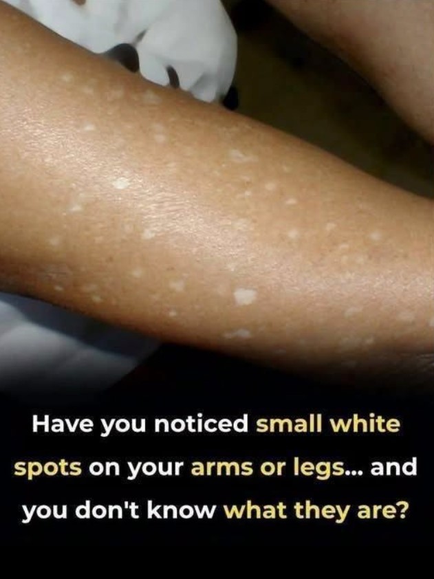
Have you ever looked down at your arms or legs and suddenly noticed several tiny white or pale spots that weren’t there before? They can be surprisingly noticeable, particularly after spending time in the sun when the surrounding skin becomes darker. For many people, the first reaction is concern: Is it an allergy? A vitamin deficiency? A fungal infection? Or something more serious?
The good news is that small, flat white spots on the arms and legs are often caused by a harmless skin condition called idiopathic guttate hypomelanosis (IGH). It is a common form of pigment loss that tends to appear on areas exposed to the sun, especially the forearms and shins. The spots are generally small, smooth, and do not cause symptoms.
However, not every white spot is IGH. Several different skin conditions can produce lighter patches, and some require specific treatment. Understanding the differences can help you know when the spots are probably harmless and when it makes sense to have them examined by a dermatologist.
What Is Idiopathic Guttate Hypomelanosis?
Idiopathic guttate hypomelanosis is a common acquired condition in which small areas of skin lose some or all of their normal pigment.
The name sounds complicated, but it can be broken down quite simply. “Idiopathic” means that there isn’t always one clearly identifiable cause, “guttate” refers to the small drop-like spots, and “hypomelanosis” means there is less melanin, the pigment responsible for much of the skin’s color.
The spots are typically flat rather than raised. They may appear pale, off-white, or porcelain-white and are often only a few millimeters across. They are particularly common on the sun-exposed portions of the forearms and lower legs.
In many cases, they don’t itch, hurt, burn, or otherwise bother the person who has them.
That is one reason people may not notice them until they look closely at their skin.
Why Do These White Spots Appear?
The exact cause of idiopathic guttate hypomelanosis isn’t completely understood.
However, the condition is associated with cumulative sun exposure and changes that occur in the skin over time. The spots are particularly common on areas that receive repeated sun exposure, such as the forearms and shins.
As skin ages and experiences years of environmental exposure, individual areas may gradually produce less pigment.
This can make the small spots stand out more dramatically, especially during summer.
For example, imagine that someone spends several weeks outdoors and develops a tan on their arms. Most of the skin becomes darker, but the small areas affected by pigment loss remain pale. The contrast can make the spots suddenly appear much more obvious.
That doesn’t necessarily mean the spots developed overnight.
They may simply have become easier to see.
What Do These Spots Usually Look Like?
IGH commonly produces multiple small, flat macules.
They can be:
- White or noticeably lighter than the surrounding skin
- Round, oval, or irregularly shaped
- Only a few millimeters wide
- Smooth rather than significantly scaly
- Scattered across the forearms or lower legs
- More obvious after tanning
- Usually painless and non-itchy
DermNet notes that the typical lesions measure around 2–5 millimeters, although some can be larger.
The spots can sometimes have unusual borders that look feathery, scalloped, or slightly irregular.
This is one reason they can look different from one person to another.
Why Do They Look Whiter After Sun Exposure?
This is one of the most confusing aspects of the condition.
The white spots themselves aren’t necessarily becoming dramatically whiter. Instead, the surrounding skin may become darker after exposure to ultraviolet light.
The result is increased contrast.
Think of a white sticker placed on a sheet of paper. Against a pale background, it might be difficult to notice. Put that same sticker against a dark background, and it immediately stands out.
A similar effect can happen with pigment-loss spots.
This is also why someone may discover the spots during the warmer months or after returning from a sunny vacation.
Is It a Sign of a Vitamin Deficiency?
Not necessarily.
It’s common for people to assume that any unusual change in skin color must be related to a nutritional deficiency. While nutrition can influence skin health, the appearance of small, stable, white macules on sun-exposed arms and legs is not enough to conclude that a person has a vitamin deficiency.
It’s better not to self-diagnose based solely on a photograph or social-media post.
If the spots are new, spreading, changing, or accompanied by other symptoms, a healthcare professional can determine whether testing is appropriate.
Could It Be Vitiligo?
Vitiligo is another important possibility when someone notices areas of lost skin pigment.
Vitiligo occurs when pigment-producing cells are damaged or destroyed, resulting in areas of depigmented skin. It can affect many parts of the body and can produce very white patches.
IGH and vitiligo can sometimes look similar, especially in photographs.
There are, however, differences that a dermatologist can evaluate. DermNet lists vitiligo among the conditions that can resemble idiopathic guttate hypomelanosis and notes that dermoscopic examination can help distinguish them.
This is why it isn’t a good idea to look at one picture online and immediately conclude that a white spot is vitiligo—or that it definitely isn’t.
Could It Be a Fungal Infection?
Another possibility is tinea versicolor, sometimes called pityriasis versicolor.
This is a superficial yeast-related skin condition that can cause lighter or darker patches. Unlike typical IGH, tinea versicolor can produce fine scaling and may sometimes itch. The patches can also gradually enlarge or merge with neighboring areas.
Tinea versicolor is especially worth considering when the spots appear on areas such as the chest, back, shoulders, or upper arms and have a subtle flaky surface.
It can also become more noticeable after tanning because the affected areas don’t tan normally.
Because treatment differs between fungal infections and pigment disorders, getting the diagnosis right is important.
What About Dry Skin?
Dry skin can sometimes make the skin look pale, flaky, or uneven.
However, ordinary dryness usually produces a different appearance from sharply defined, smooth white macules.
If the skin is visibly flaky, rough, cracked, itchy, or irritated, dryness or another inflammatory skin condition may be contributing to the appearance.
The distinction matters because treating dryness is very different from treating a disorder involving pigment.
What About Keratosis Pilaris?
Keratosis pilaris is another extremely common skin condition that can create tiny bumps on the arms and legs.
But there is an important difference.
Keratosis pilaris generally creates small raised, rough-feeling bumps, rather than flat white patches. The American Academy of Dermatology describes them as plugs of dead skin cells that commonly appear on the upper arms and thighs.
If you run your fingers over the affected area and it feels like fine sandpaper, keratosis pilaris may be more likely than a flat pigment disorder.
Do These White Spots Need Treatment?
If the spots are diagnosed as idiopathic guttate hypomelanosis, treatment is often unnecessary from a medical standpoint.
DermNet describes IGH as a benign condition and notes that reassurance is generally all that is needed. Sun protection is recommended as well.
Some people, however, dislike the appearance of the spots and want to explore cosmetic treatment.
Dermatologists may consider options such as cryotherapy, certain topical treatments, chemical peels, laser-based procedures, or other approaches depending on the individual situation.
These treatments aren’t something to experiment with at home.
A treatment that is appropriate for one pigment disorder could be ineffective—or potentially irritating—for another.
Sun Protection Still Matters
Even when the white spots themselves are harmless, protecting your skin from excessive ultraviolet exposure is important.
Use broad-spectrum sunscreen, wear protective clothing when appropriate, and seek shade during periods of intense sun.
Sun protection won’t necessarily make existing IGH spots disappear, but it can help reduce additional sun damage and limit the contrast between the spots and surrounding tanned skin.
It is also a good general habit for maintaining healthier skin over time.
When Should You See a Dermatologist?
Most small, stable, symptom-free pale spots aren’t an emergency.
Still, there are situations in which getting professional advice is worthwhile.
Consider making an appointment if:
- The spots are rapidly increasing in number
- Existing spots are becoming significantly larger
- They are spreading to many new areas
- The skin becomes itchy, painful, inflamed, or scaly
- The patches are dramatically different from the surrounding skin
- You notice pigment changes elsewhere on your body
- You’re unsure whether the spots are white, pale, or actually scaly
- You’re concerned that the changes could represent vitiligo or another skin disorder
A dermatologist can usually learn a great deal simply by examining the skin. In some cases, additional tools such as a Wood lamp or dermoscopy may help distinguish different pigment disorders.
Don’t Scrub or Pick at the Spots
One common mistake is assuming that a white spot must be something sitting on top of the skin.
If the change is caused by pigment loss, scrubbing won’t restore the color.
Aggressive exfoliation can instead irritate the skin and make the area more noticeable.
Avoid harsh scrubbing, picking, bleaching products, or unproven home remedies simply because you saw them recommended online.
When you’re dealing with unexplained changes in pigmentation, gentle skin care is usually a better approach until you know what you’re actually treating.
Pay Attention to the Pattern
One useful clue is where the spots appear.
IGH has a strong tendency to affect sun-exposed areas, particularly the forearms and shins.
If the spots are concentrated in these locations, are small and flat, and have remained relatively stable without itching or scaling, IGH may be one possibility.
On the other hand, if the spots appear in unusual locations, are accompanied by scaling, or form larger patches, another diagnosis may be more appropriate.
The overall pattern matters more than one isolated spot.
Why These Spots Can Look More Alarming Than They Are
Skin changes naturally attract attention.
A new mark can make someone immediately imagine the worst, particularly when it involves a visible change in color.
But a photograph doesn’t provide enough information to diagnose a skin condition.
Lighting, camera exposure, skin tone, tanning, dryness, and even the angle of the photograph can dramatically change how a spot looks.
That’s why the image accompanying this topic should be viewed as an example of what small white spots can look like—not as proof that every person with similar-looking spots has the same condition.
What You Can Do at Home
If your spots are mild and you have no concerning symptoms, focus on basic skin care while arranging professional evaluation if you’re uncertain.
Use a gentle cleanser, moisturize regularly, avoid unnecessary irritation, and protect exposed skin from excessive sunlight.
Don’t attempt to remove the spots mechanically.
If you notice changes over time, taking occasional photographs under similar lighting can also help you and your healthcare professional compare their appearance.
The Bottom Line
Seeing small white spots on your arms or legs can certainly be unsettling, especially when they seem to appear out of nowhere. But in many cases, these spots are associated with idiopathic guttate hypomelanosis, a common and benign pigment condition that frequently affects sun-exposed areas such as the forearms and shins.
The spots are typically small, flat, and symptom-free, and they may become much more noticeable after the surrounding skin tans. They don’t necessarily indicate a serious disease, and when IGH is confirmed, treatment often isn’t medically necessary.
At the same time, it’s important not to assume that every white spot is IGH. Vitiligo, tinea versicolor, post-inflammatory pigment changes, and other skin conditions can produce similar-looking changes. Tinea versicolor, for example, may cause lighter patches with fine scaling, while vitiligo can produce areas of more complete pigment loss.
So if you’ve noticed these spots, don’t panic—but don’t try to diagnose yourself from a picture either. Pay attention to their size, shape, location, texture, and whether they’re changing. Protect your skin from excessive sun exposure, avoid harsh home treatments, and see a dermatologist if the appearance is new, spreading, symptomatic, or simply concerning to you.
Sometimes the most reassuring answer is also the simplest one: a surprising change in the appearance of your skin doesn’t automatically mean something is seriously wrong. Getting the right diagnosis can replace uncertainty with peace of mind.
Conclusion :
Small white spots on the arms or legs can look alarming, but they are not always a sign of something serious. One possible cause is idiopathic guttate hypomelanosis (IGH), a common skin condition that appears as small, smooth, lighter-colored spots, particularly on areas that receive regular sun exposure. Other conditions, including fungal infections, vitiligo, post-inflammatory changes, or other pigment disorders, can sometimes look similar.
Because the appearance of these spots can vary from person to person, it is best not to diagnose them based on a photo alone. If the spots are spreading, changing in appearance, becoming itchy or painful, or appearing suddenly, consider having them evaluated by a dermatologist. A professional can determine the cause and recommend the appropriate treatment if one is needed.
Most importantly, noticing changes in your skin is a good reason to pay attention rather than panic. Protecting your skin from excessive sun exposure and keeping track of any new or changing spots can help you better understand what is happening. When in doubt, a medical professional can provide reassurance and an accurate diagnosis.