Finding a strange new growth on a loved one’s skin can be surprisingly alarming. It becomes even more concerning when the spot seems to have appeared quickly, looks noticeably different from the surrounding skin, and has a hard or crusty center.
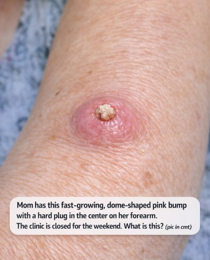
That is exactly what makes the bump in this photograph so attention-grabbing. It appears as a raised, pink, rounded lesion on the forearm with a prominent firm-looking plug in the middle. According to the description, it has also been growing rapidly.
One possibility that dermatologists would consider when seeing this particular pattern is a keratoacanthoma, often abbreviated as KA. Keratoacanthomas are distinctive skin growths that can develop relatively quickly and may have a classic dome or crater-like shape with a central accumulation of keratin. They are commonly found on areas exposed to sunlight, including the arms and face.
But there is an important complication.
A lesion that looks like a keratoacanthoma can closely resemble cutaneous squamous cell carcinoma (SCC), a form of skin cancer. Because of that overlap, it would be unsafe to look at the photograph and declare, “That’s definitely a keratoacanthoma.” A healthcare professional may need to examine the lesion directly and, depending on its appearance, perform a biopsy or remove it for laboratory examination.
So what should someone do if this happens over the weekend, when the usual clinic isn’t open?
The answer is not to panic—but it is also not to ignore the lesion for weeks.
The safest approach is to leave the bump alone, avoid trying to remove the hard center at home, protect it from irritation, document how it looks and changes, and arrange an appropriate medical evaluation as soon as possible.
This article takes a closer look at what a bump like this might represent, why its appearance is important, what keratoacanthoma means, how it differs from other skin growths, why doctors take these lesions seriously, what usually happens at an appointment, and which warning signs should make someone seek more urgent care.
Important: A photograph cannot establish a diagnosis. The information below is educational and is not a substitute for an examination by a dermatologist or other qualified healthcare professional.
1. What Are We Looking at in the Photograph?
The first thing that stands out is the overall shape.
Rather than looking like a flat discoloration or a small ordinary mole, the lesion appears to be a raised, rounded bump. The surrounding skin has a pink-to-red appearance, while the middle contains a lighter, rough-looking material.
That central material is particularly interesting because keratin can accumulate in certain skin lesions and form a hard, crusty or horn-like plug.
The combination of:
- rapid growth,
- a rounded or dome-shaped profile,
- pink or reddish coloration,
- firmness,
- and a central keratin-like plug
is one reason a dermatologist might consider keratoacanthoma among the possibilities.
DermNet describes keratoacanthomas as typically being solitary, rapidly growing nodules on sun-exposed skin, often involving the face or upper limbs. The classic lesion is firm, sharply defined, red or skin-colored, and has a central hyperkeratotic plug.
However, these characteristics are clues rather than proof.
Many skin conditions can overlap in appearance, and even experienced clinicians sometimes need tissue examination before they can confidently distinguish one lesion from another.
That distinction is particularly important here because one of the main conditions that can resemble keratoacanthoma is squamous cell carcinoma.
2. What Is a Keratoacanthoma?
A keratoacanthoma is a distinctive skin tumor that generally develops relatively quickly.
It is often described as a dome-shaped or crateriform growth containing a central plug of keratin. Some lesions begin as a small bump and enlarge noticeably over a period of weeks. They may eventually stabilize and, in some cases, regress, leaving a scar.
The name itself gives us a clue about its appearance.
“Kerato” refers to keratin, a tough protein that forms much of the outer layer of the skin, hair, and nails. “Acanthoma” refers to a particular type of skin growth.
The central material seen in a classic keratoacanthoma is therefore not necessarily pus or a foreign object. It can represent a dense accumulation of keratin.
This is one reason people may be tempted to squeeze or pick at the center.
It can look almost like a plug that should simply come out.
But that is exactly what should not be done at home.
Trying to pull, cut, scrape, burn, freeze, or squeeze a suspicious skin growth can cause bleeding and inflammation, damage surrounding tissue, introduce infection, and make the lesion more difficult for a clinician to assess.
More importantly, removing the visible surface material does not answer the bigger question: what caused the growth in the first place?
3. Why Does It Look Like a Tiny Volcano?
People sometimes describe a classic keratoacanthoma as looking like a tiny volcano or crater.
That description comes from its architecture.
The lesion can develop a raised outer border surrounding a central area filled with compacted keratin. Viewed from the side, the growth may look like a dome. Viewed from above, the central plug can make it resemble a small crater.
DermNet describes the classic lesion as having a central hyperkeratotic plug and an even shoulder around the center.
This is quite different from many common skin marks.
A typical freckle, for example, is flat.
A simple age spot is generally flat.
A small mole may be raised, but it usually doesn’t have this particular combination of rapid enlargement and central keratin accumulation.
A wart can also be raised and rough, which is why a wart may enter the differential diagnosis, but the overall pattern and history can be different.
A cyst may have a central opening or pore, but its texture, growth pattern, and underlying structure can differ.
The appearance therefore provides useful information—but not enough information for a final diagnosis.
4. Why Is the Central Plug So Important?
The central plug is one of the most recognizable features associated with keratoacanthoma.
Keratin is continuously produced by the skin. Normally, old skin cells shed naturally. In certain lesions, however, keratin can become densely packed within a growth.
This can produce a hard, pale, yellowish, tan, or crust-like center.
In the photograph, the central area appears much lighter than the pink skin surrounding it.
That visual contrast can make the lesion especially noticeable.
But a central crust or plug is not exclusive to keratoacanthoma.
Other lesions—including some forms of skin cancer—can become crusted, keratinized, or ulcerated.
The American Academy of Dermatology notes that squamous cell carcinoma can appear as a firm, dome-shaped growth, as well as a rough patch, sore, wart-like growth, or horn-like growth.
That is why a hard center should be treated as a reason for examination rather than an invitation to dig it out.
5. Why Is Rapid Growth Such a Big Deal?
The word “fast-growing” deserves attention.
Many ordinary skin changes develop slowly.
A spot might remain nearly identical for months or years.
A rapidly enlarging lesion behaves differently.
Keratoacanthomas are notable because they can grow considerably over a relatively short period. DermNet describes solitary keratoacanthomas as capable of growing rapidly over several weeks, sometimes reaching roughly 1–2 centimeters before later stabilizing or regressing.
This rapid evolution is one of the reasons the lesion can seem to appear “out of nowhere.”
Someone might remember that the area looked normal a month ago and then suddenly notice a prominent bump.
However, rapid growth does not automatically mean cancer.
It simply means that the lesion deserves closer attention.
The American Academy of Dermatology also lists rapid growth among possible features of squamous cell carcinoma, which is another reason doctors take a quickly enlarging lesion seriously.
6. Why the Forearm Is a Relevant Location
The forearm is a common area of sun exposure.
People may expose their arms to ultraviolet radiation without thinking about it.
Driving, gardening, walking, sitting outside, doing outdoor chores, exercising, traveling, or simply spending time outdoors can all contribute to cumulative UV exposure.
The issue is not just dramatic sunburns.
Skin receives ultraviolet exposure repeatedly over a lifetime.
Keratoacanthomas are commonly found on sun-exposed areas, including the upper limbs and face. DermNet identifies ultraviolet exposure as one of the important risk factors associated with these lesions.
Squamous cell carcinoma is also strongly associated with cumulative ultraviolet exposure.
This doesn’t mean that every forearm bump is caused by sunlight.
It means the location is one piece of the puzzle that a dermatologist may consider alongside age, skin type, medical history, previous skin lesions, medications, immune status, and the behavior of the growth.
7. Could Sun Exposure Really Matter Years Later?
Yes.
One reason skin examination becomes increasingly important with age is that ultraviolet exposure accumulates over time.
Someone may not remember every summer spent outdoors as a child or every afternoon working in a garden decades ago.
The skin, however, does not forget ultraviolet exposure in the same way.
Cumulative UV damage is an important risk factor for several types of skin cancer.
That is why dermatologists recommend protecting exposed skin even on ordinary days rather than thinking sunscreen is only necessary for beach vacations.
The American Academy of Dermatology recommends broad-spectrum, water-resistant sunscreen with an SPF of 30 or higher, along with shade and protective clothing.
8. Is Keratoacanthoma Actually Cancer?
This is where the terminology becomes confusing.
Keratoacanthoma has traditionally been described as a benign or low-grade skin tumor, and some lesions can regress spontaneously. But clinically, it can be extremely difficult to distinguish from well-differentiated squamous cell carcinoma.
Because of this overlap, dermatologists and pathologists may use terminology such as “squamous cell carcinoma, keratoacanthoma type”.
DermNet specifically notes that many clinicians and pathologists prefer to consider these lesions within the SCC spectrum and recommend surgical removal because the clinical course can be unpredictable.
This is why simply searching for photographs and deciding that a lesion “looks like a keratoacanthoma” isn’t enough.
The important question isn’t only:
“Does this look like a keratoacanthoma?”
The important question is:
“Can we confidently exclude a squamous cell carcinoma or another concerning lesion?”
That may require tissue.
9. What Is Squamous Cell Carcinoma?
Cutaneous squamous cell carcinoma is a common form of skin cancer that develops from abnormal squamous cells in the skin.
It can appear in several different ways.
According to the American Academy of Dermatology, it may look like:
- a rough or scaly patch,
- a firm dome-shaped growth,
- a sore surrounded by raised skin,
- a sore that does not heal,
- a wart-like growth,
- or a horn-like growth.
Some lesions may itch, hurt, burn, bleed, become crusted, or feel tender.
Others may cause very little discomfort.
That last point is important.
The absence of pain does not automatically mean that a skin growth is harmless.
People sometimes assume that a dangerous lesion must hurt.
That isn’t necessarily true.
A suspicious skin growth may be completely painless, particularly early in its development.
10. Why Squamous Cell Carcinoma Can Be Confused With Keratoacanthoma
The two can share several characteristics.
Both can:
- grow relatively quickly,
- appear as raised nodules,
- occur on sun-exposed skin,
- become keratinized,
- form a central crater or plug,
- appear pink or red,
- and occur in older adults.
That overlap is precisely what makes diagnosis challenging.
DermNet states that keratoacanthomas may be clinically indistinguishable from well-differentiated SCC and that even dermoscopy cannot reliably separate the two in every case.
This is why the appropriate response to an image like this is not panic and not complacency.
It is evaluation.
11. Does This Photograph Prove It Is Cancer?
No.
And it would be irresponsible to say that it does.
A photograph can show visible features, but it cannot reveal everything happening underneath the surface.
A dermatologist can evaluate:
- how firm the lesion is,
- whether the borders are well defined,
- whether it is attached to deeper tissue,
- whether there are additional lesions nearby,
- how the surrounding skin feels,
- whether lymph nodes are enlarged when clinically relevant,
- and whether a biopsy is appropriate.
Laboratory examination of tissue can provide information that a photograph simply cannot.
The American Academy of Dermatology explains that a skin biopsy is needed to establish a diagnosis of skin cancer such as SCC.
So the photograph should be considered a visual clue, not a diagnosis.
12. What Other Things Could Look Like This?
Although keratoacanthoma and squamous cell carcinoma are important possibilities, they are not the only ones.
DermNet lists several conditions in the differential diagnosis of keratoacanthoma, including:
- well-differentiated SCC,
- nodular basal cell carcinoma,
- common warts,
- certain forms of melanoma,
- giant molluscum contagiosum,
- nodular prurigo,
- and other less common lesions.
A clinician may also consider an inflamed cyst or other growth depending on the history and physical examination.
This is another reason not to become fixated on one diagnosis after looking at a photograph.
The same general visual pattern can arise from different causes.
13. Could It Be a Wart?
A wart can certainly be raised and rough.
Viral warts often have an uneven or thickened surface and can occur on various parts of the body.
But a classic keratoacanthoma tends to have a different overall architecture: a rapidly growing, firm nodule with a central keratin plug and relatively smooth or sharply defined surrounding shoulder.
The history matters.
A wart often develops differently and may remain for a longer period.
But there are exceptions.
That is why the clinician doesn’t diagnose solely from one visual characteristic.
14. Could It Be a Cyst?
An inflamed cyst can also produce a raised, red bump.
Some cysts have a visible opening or central point, which might look superficially similar to the center of the lesion in the photograph.
However, a cyst and a keratoacanthoma are structurally different conditions.
A cyst is generally a sac-like structure containing material such as keratin.
A keratoacanthoma is a tumor-like proliferation of skin cells with a distinctive architecture.
The growth rate and appearance can help the clinician distinguish between them.
Again, squeezing the lesion isn’t a good idea.
Even if it were a cyst, manipulating it can cause inflammation, rupture, bleeding, or infection.
15. Could It Be an Ingrown Hair?
On certain parts of the body, an ingrown hair can produce a red bump.
But the forearm lesion shown here does not immediately suggest the typical appearance of a simple ingrown hair, particularly given the described rapid enlargement and firm central plug.
Still, visual resemblance isn’t enough to establish a diagnosis.
If the bump is new, growing, and unusual, professional evaluation remains the safest option.
16. Could It Be an Infection?
Infections can cause red, swollen, tender bumps.
An abscess may become painful and contain pus.
Cellulitis can produce spreading redness and warmth.
But the photograph’s central hard, keratin-like material is different from the typical appearance of liquid pus.
More importantly, the clinical history matters.
If the surrounding redness is rapidly expanding, the area becomes very warm or swollen, severe pain develops, pus appears, or fever occurs, infection becomes a more urgent concern.
The situation then changes from “schedule a dermatology evaluation” to “seek prompt medical care.”
17. What Should Mom Do While the Clinic Is Closed?
If the lesion is stable and Mom otherwise feels well, there is usually no reason to panic simply because the clinic is closed for the weekend.
But there is also no good reason to spend several weeks waiting to see whether it disappears.
A reasonable short-term plan is:
Leave it alone.
Do not squeeze it.
Do not scratch it.
Do not try to remove the central plug.
Do not cut it.
Do not burn it.
Do not freeze it with an at-home device.
Do not apply strong wart-removal products.
Do not repeatedly scrape the surface.
Instead, keep the area clean and protected from unnecessary friction.
If clothing repeatedly rubs against it, a clean non-stick dressing can help protect the lesion until it can be evaluated.
18. Why Picking at the Plug Is a Bad Idea
The center can look extremely tempting.
It may appear as though there is something trapped inside that needs to come out.
But the central keratin is part of the lesion’s structure.
Removing the visible material doesn’t remove the underlying growth.
Picking can cause:
- bleeding,
- pain,
- inflammation,
- bacterial contamination,
- delayed healing,
- additional scarring,
- and difficulty interpreting the lesion.
It can also change the appearance that a dermatologist needs to see.
So if the central plug looks loose or crusty, resist the urge to pull it off.
Let the healthcare professional decide what needs to be removed and how.
19. What About Home Wart Removers?
Home wart treatments may contain acids such as salicylic acid.
Those products are designed for specific conditions and should not automatically be used on an undiagnosed growth.
Applying an acid to a suspicious lesion can irritate or damage the surrounding skin and alter the appearance of the lesion.
More importantly, if the growth isn’t a wart, treating it as one could delay the correct diagnosis.
A rapidly growing dome-shaped lesion with a central keratin plug is not the kind of skin change that should be experimentally treated at home.
20. Should It Be Covered?
If the bump is not being irritated, it may not need a dressing.
But if sleeves, bedding, furniture, or other objects repeatedly rub against it, protecting the area can be sensible.
A clean, non-stick dressing can reduce friction.
The goal isn’t to “treat” the growth.
The goal is simply to prevent unnecessary trauma while waiting for professional evaluation.
The area should be kept reasonably clean, and the dressing should be changed if it becomes wet or dirty.
21. Should You Take Photographs?
Yes, photographs can be useful for documenting changes.
But they should supplement—not replace—medical evaluation.
Take a clear photograph in good lighting.
Try to keep the distance and angle consistent.
If measuring the lesion, place a ruler beside it rather than pressing the ruler against the bump.
A daily photograph may be enough to document short-term changes.
There is no need to photograph it dozens of times a day.
What matters most is a useful record showing whether it is getting larger, changing color, bleeding, crusting, or becoming more inflamed.
22. What Information Should Be Written Down?
Before the appointment, it can help to make a short timeline.
Write down:
When was the bump first noticed?
Was it there for months, or did it seem to appear recently?
How quickly did it grow?
Even an approximate timeline is useful.
Has the color changed?
For example, was it initially skin-colored and later pink or red?
Has the center changed?
Did a crust or hard plug appear later?
Does it hurt?
Or is it completely painless?
Does it itch or sting?
Has it bled?
Has it been repeatedly traumatized?
Has Mom had similar lesions before?
Has she ever had skin cancer or precancerous lesions?
Does she take medications that suppress the immune system?
These details can help the clinician understand the lesion’s behavior.
23. What Happens at the Dermatologist’s Office?
The first step is generally a visual and physical examination.
The clinician may examine the lesion using magnification or a dermatoscope.
They may ask questions about:
- when it appeared,
- how quickly it changed,
- whether it bleeds,
- whether it hurts,
- previous skin cancers,
- sun exposure,
- medications,
- immune status,
- and other relevant medical history.
The lesion may also be measured.
The clinician will look at the borders, color, surface, central material, and overall structure.
But even a highly experienced dermatologist may not be able to make a definitive diagnosis simply by looking.
24. Why a Biopsy May Be Needed
A biopsy involves removing a portion or all of a lesion so that the tissue can be examined under a microscope.
The area is usually numbed with a local anesthetic.
The clinician then removes tissue using the technique most appropriate for the lesion.
The specimen is sent to a pathology laboratory.
A dermatopathologist or other trained pathologist examines the cells and architecture.
This microscopic examination can help determine whether the lesion is:
- benign,
- keratoacanthoma-like,
- squamous cell carcinoma,
- or another condition.
The American Academy of Dermatology states that a skin biopsy is the only way to know whether a suspicious lesion is skin cancer.
25. Why the Type of Biopsy Matters
Not every biopsy provides the same amount of information.
A superficial sample may not capture the entire architecture of a lesion.
DermNet specifically notes that partial or shave samples may sometimes be inadequate for confidently assessing keratoacanthoma because the relevant features can involve the deeper or peripheral portions of the lesion.
That doesn’t mean every patient needs a particular type of biopsy.
The clinician decides based on the lesion’s size, location, appearance, suspected diagnosis, and other factors.
The important point is that the method should be selected by the healthcare professional rather than improvised at home.
26. What If the Doctor Recommends Removing the Whole Lesion?
That can be a very reasonable approach.
Because keratoacanthoma and SCC can overlap clinically, complete removal can both treat the lesion and provide tissue for definitive pathological examination.
DermNet notes that most keratoacanthomas are treated surgically and that excision is generally prudent because of the difficulty distinguishing them from SCC.
The exact procedure depends on the individual case.
A small lesion on the forearm may be removed relatively straightforwardly under local anesthesia.
The area can then be closed or allowed to heal according to the technique used.
27. Does Removal Hurt?
Most minor skin procedures are performed after numbing the area.
The injection of local anesthetic can sting briefly.
Once the area is numb, the patient should generally feel pressure or movement rather than sharp pain.
Afterward, there may be some soreness, tenderness, bruising, or tightness depending on the procedure.
The healthcare team will provide wound-care instructions.
Patients should follow those instructions rather than applying homemade remedies.
28. What If the Pathology Says Squamous Cell Carcinoma?
Hearing the words “skin cancer” can understandably be frightening.
But cutaneous squamous cell carcinoma is highly treatable when detected early.
The American Academy of Dermatology states that early SCC is highly treatable and that more than 90% of patients are cured.
Treatment depends on factors such as:
- tumor size,
- depth,
- location,
- pathology findings,
- whether it has high-risk characteristics,
- whether it has spread,
- and the patient’s overall health.
For many localized lesions, surgical treatment is the main approach.
29. What Is Standard Excision?
Standard surgical excision involves numbing the area and removing the visible tumor along with a margin of surrounding skin.
The removed tissue is sent to a laboratory.
The pathology report can determine whether the cancer has been completely removed and whether additional treatment is needed.
The American Academy of Dermatology explains that surgical excision is widely used for SCC that remains confined to the skin and has a lower risk of spreading or returning.
30. What Is Mohs Surgery?
Mohs micrographic surgery is a specialized technique designed to remove skin cancer while preserving as much healthy tissue as possible.
It is particularly useful for selected tumors in areas where tissue conservation matters or where the cancer has characteristics that make precise margin control especially important.
Not every lesion requires Mohs surgery.
A dermatologist or Mohs surgeon determines whether it is appropriate based on the specific cancer and its location.
31. Does a Diagnosis of SCC Mean It Has Spread?
No.
Most cutaneous squamous cell carcinomas are detected while still localized.
The American Academy of Dermatology emphasizes that early SCC is highly treatable.
The word “cancer” can cause people to immediately imagine widespread disease.
But skin cancer is not automatically advanced cancer.
The stage and risk level depend on the individual tumor.
That is why early evaluation is valuable.
32. What Happens If a Keratoacanthoma Regresses on Its Own?
This is another confusing part of the story.
Classic keratoacanthoma has historically been described as having a pattern of rapid growth followed by stabilization and eventual regression.
DermNet reports that a typical solitary lesion may eventually regress over several months and leave a depressed scar.
That sounds reassuring.
But it does not mean that people should wait months for a suspicious lesion to disappear.
The problem is that the clinician may not be able to determine with confidence whether the lesion is a regressing keratoacanthoma or an SCC with a similar appearance.
Therefore, medical evaluation remains important even when spontaneous regression is theoretically possible.
33. Why “It Might Go Away” Isn’t a Good Reason to Ignore It
Imagine seeing a bump that grows rapidly.
Someone might say:
“It could be a keratoacanthoma, and those can disappear on their own.”
The problem is that the same lesion might instead represent squamous cell carcinoma.
Waiting to see what happens can therefore create unnecessary risk.
DermNet recommends treating a KA-like lesion as SCC until proven otherwise in appropriate circumstances.
That doesn’t mean every lesion is cancer.
It means the consequences of overlooking SCC are important enough that doctors generally prefer a definitive evaluation.
34. What Risk Factors Make Evaluation Especially Important?
Certain factors may increase concern.
These include:
- older age,
- substantial lifetime sun exposure,
- previous skin cancer,
- multiple precancerous lesions,
- immune suppression,
- organ transplantation,
- certain medications,
- a history of radiation to the area,
- and certain genetic conditions.
DermNet identifies chronic UV exposure and immune suppression among important associations with keratoacanthoma.
However, the absence of these risk factors does not make a rapidly growing lesion automatically harmless.
A suspicious lesion deserves evaluation based on its own behavior.
35. What If Mom Has Never Had Skin Problems Before?
That is reassuring in one sense, but it doesn’t eliminate the need to check the bump.
Skin cancers and other unusual growths can occur in people who have never previously had a suspicious lesion.
A new lesion that behaves differently from the surrounding skin is worth discussing with a clinician.
The key principle is:
Don’t compare the lesion only to someone else’s risk profile. Compare it to its own behavior.
If it is new and changing, that matters.
36. What If It Doesn’t Hurt?
Again, that doesn’t settle the question.
Some suspicious skin lesions cause pain or tenderness.
Others don’t.
The American Academy of Dermatology lists pain, tenderness, itching, numbness, stinging, and burning as possible symptoms of SCC, but it also notes that some people may simply notice a growth or change in their skin.
So a painless bump can still warrant examination.
37. What If It Starts Bleeding?
Bleeding is an important change to document.
A lesion can bleed because it has been scratched, bumped, or irritated by clothing.
But repeated spontaneous bleeding is also a reason to have a skin growth examined.
If bleeding is minor, applying gentle direct pressure with clean gauze can help.
If bleeding is heavy or does not stop after sustained pressure, urgent medical assessment may be appropriate.
The key is not to keep picking at a bleeding lesion to “see what happens.”
38. What If the Area Becomes Redder?
Some redness can occur around a skin growth.
But rapidly spreading redness is different.
If the redness expands outward, the area becomes increasingly warm and swollen, pus appears, severe pain develops, or Mom develops fever or chills, infection becomes a concern.
In that situation, the appropriate response may be urgent medical care rather than waiting for a routine dermatology appointment.
39. What If the Bump Doubles in Size Over a Few Days?
Very rapid change deserves attention.
A lesion that suddenly becomes much larger, dramatically more painful, or begins bleeding heavily should be assessed promptly.
The exact urgency depends on the circumstances.
A stable lesion that has slowly increased over several weeks is different from a lesion that is rapidly changing over hours or days with severe inflammation.
When in doubt, contacting an urgent-care service or healthcare professional can help determine whether waiting is appropriate.
40. When Is It Reasonable to Wait Until Monday?
If the clinic is closed for the weekend and Mom:
- feels generally well,
- has no severe pain,
- has no rapidly spreading redness,
- has no significant swelling,
- has no fever,
- and the lesion is not bleeding uncontrollably,
then arranging the earliest appropriate appointment when the clinic reopens may be reasonable.
The important distinction is between short-term waiting for an appointment and long-term ignoring of the lesion.
Waiting two days for a clinic to reopen is not the same thing as watching a suspicious lesion for two months.
41. When Should You Not Wait?
Seek more urgent evaluation if there is:
- uncontrolled bleeding,
- severe or rapidly increasing pain,
- rapidly spreading redness,
- significant swelling,
- pus accompanied by worsening inflammation,
- fever or systemic illness,
- rapid dramatic expansion,
- involvement of an area where function may be affected,
- or other concerning changes.
People who are significantly immunocompromised or have a history of aggressive skin cancer may also need a lower threshold for seeking prompt evaluation.
The exact recommendation should be based on the individual’s medical situation.
42. Why the Weekend Situation Can Cause Extra Anxiety
A strange skin lesion discovered on a Friday evening can feel much scarier than the same lesion discovered on a Monday morning.
The reason is simple: access to reassurance is temporarily limited.
People start searching through photographs online.
They compare the bump with images.
They imagine the worst possibility.
Then they wonder whether they should go to an emergency department.
This is understandable.
But internet photographs are not a substitute for clinical assessment.
The goal over the weekend should be to protect the lesion and watch for meaningful changes, not to repeatedly diagnose it from photographs.
43. Don’t Let Internet Images Decide the Diagnosis
Searching images can be useful for learning general concepts.
It is not reliable for identifying a specific lesion.
Two unrelated skin conditions can look nearly identical in a photograph.
Lighting can change color.
Camera focus can change apparent texture.
Skin tone can affect how redness appears.
The photograph may also fail to show the lesion’s depth or firmness.
A dermatologist has the advantage of seeing, touching, measuring, and examining the lesion directly.
And if necessary, pathology provides information that no photograph can provide.
44. Why the Image Is Still Useful
That doesn’t mean the photograph has no value.
Quite the opposite.
A good photograph can help document how the lesion looked at a particular point in time.
If it changes before the appointment, comparison photographs may help the clinician understand the timeline.
The photograph can also help a healthcare professional see what initially caught the patient’s attention.
But it should be treated as documentation—not diagnosis.
45. How to Take a Better Tracking Photo
If you need to document the lesion:
- Use good natural light.
- Keep the camera approximately the same distance away each time.
- Take one overall photograph showing where the lesion is located.
- Take one closer photograph showing its surface.
- If measuring it, place a ruler beside it without pressing into the skin.
- Avoid filters.
- Avoid heavy digital zoom.
- Record the date.
- Note symptoms such as tenderness or bleeding.
This creates a simple visual record that can be much more useful than dozens of random photographs.
46. Why You Shouldn’t Keep Touching It
Repeated touching can irritate the lesion.
Fingernails can introduce bacteria.
Rubbing can remove crust and cause bleeding.
And constant manipulation makes it difficult to know whether the lesion itself is changing or whether the person is changing it through repeated trauma.
The best approach is surprisingly boring:
Look, document, protect, and leave it alone.
47. What Does “Keratin Plug” Actually Mean?
Keratin is a structural protein.
It is abundant in the outer layer of the skin, hair, and nails.
When keratin accumulates within a skin lesion, it can become thick, compact, and hard.
That compact material can appear white, yellow, tan, brownish, or gray depending on the lesion and surface changes.
In keratoacanthoma, the central hyperkeratotic plug is one of the classic clinical characteristics.
So the center in the photograph isn’t necessarily a foreign object.
It may be part of the lesion itself.
48. Why Removing the Plug Doesn’t Solve the Problem
Suppose someone sees the central material and thinks:
“If I can just pull that out, maybe the bump will go away.”
Unfortunately, that’s not how these lesions work.
The central keratin is produced within the growth.
Removing surface material doesn’t necessarily remove the cells responsible for the lesion.
Worse, traumatizing the area can create bleeding and inflammation.
The correct question is not “How do I get the plug out?”
It’s:
“What is producing this plug, and does the lesion need to be biopsied or removed?”
That is the question a healthcare professional can answer.
49. What Does “Locally Destructive” Mean?
DermNet describes keratoacanthoma as a rapidly growing, locally destructive skin tumor.
That phrase can sound frightening.
It doesn’t necessarily mean the lesion has spread throughout the body.
“Locally” means the lesion can damage or disrupt nearby skin and tissue as it grows.
A growing mass can stretch, ulcerate, bleed, or destroy nearby structures.
This is another reason doctors don’t necessarily recommend simply waiting indefinitely for regression.
50. Can Keratoacanthoma Leave a Scar?
Yes.
Even when a keratoacanthoma regresses, it can leave a depressed scar.
DermNet describes spontaneous regression followed by scarring in typical solitary lesions.
Surgical treatment can also leave a scar.
The final appearance depends on the lesion, procedure, location, wound healing, and individual factors.
When treating a suspicious growth, however, the priority is first to establish an accurate diagnosis and remove or treat the lesion appropriately.
Cosmetic considerations can then be incorporated into the treatment plan.
51. Is a Scar Better Than Ignoring a Suspicious Lesion?
When a lesion is potentially cancerous, diagnosis and appropriate treatment come first.
A small scar from a properly performed procedure is generally a much more manageable problem than allowing a skin cancer to grow untreated.
That is one reason dermatologists may recommend removal even when a lesion resembles a potentially self-regressing keratoacanthoma.
52. What Happens After a Biopsy?
After the procedure, the tissue is processed by a pathology laboratory.
The pathologist examines the cells and tissue architecture.
The report may identify a benign lesion, a keratoacanthoma-like lesion, SCC, or another diagnosis.
The doctor then uses that information to determine whether additional treatment is needed.
The American Academy of Dermatology notes that pathology reports can provide information about the type of cancer, how deeply it has grown, and whether high-risk features are present.
53. Why the Pathology Report Matters So Much
A pathology report can answer questions that the photograph cannot.
It can help determine:
- what kind of cells are present,
- whether the cells are abnormal,
- whether the lesion has invaded surrounding tissue,
- and whether additional treatment may be appropriate.
That is why getting a professional evaluation is much more useful than trying to match the lesion to a photograph online.
54. What If the Result Is Benign?
That would be good news.
Not every suspicious bump is cancer.
A benign diagnosis can provide reassurance and may mean that no additional treatment is necessary beyond what has already been performed.
But even after a benign diagnosis, the experience can be a useful reminder to pay attention to future skin changes.
55. What If the Result Is Keratoacanthoma?
If pathology supports keratoacanthoma, the clinician will consider whether the lesion has already been adequately removed.
Depending on the exact pathology and procedure, additional treatment may or may not be necessary.
The management plan is individualized.
The important thing is that the lesion has been identified rather than guessed at.
56. What If It Is Squamous Cell Carcinoma?
If it is SCC, the dermatologist will determine its risk category and whether additional treatment is necessary.
Many early SCCs can be treated successfully with local treatment.
The American Academy of Dermatology states that surgery is commonly preferred for many early skin cancers and that SCC found early is highly treatable.
The earlier an appropriate diagnosis is made, the more options the clinician may have.
57. Why Early Detection Matters
Skin cancers can become more difficult to treat as they grow deeper or spread.
The AAD notes that waiting to obtain care gives SCC time to grow, potentially making treatment more difficult.
That doesn’t mean someone should panic because a bump was discovered over the weekend.
It means that once a suspicious lesion has been recognized, it should not be ignored indefinitely.
58. What About Other Skin Cancers?
Basal cell carcinoma and melanoma can also occur on sun-exposed skin.
They don’t necessarily look like this photograph.
But because skin tumors can have unusual appearances, doctors consider multiple possibilities when evaluating a new or changing lesion.
DermNet includes nodular basal cell carcinoma and amelanotic melanoma among the differential diagnoses for keratoacanthoma-like lesions.
This is another reason why a dermatologist may prefer tissue diagnosis when the appearance is uncertain.
59. Does the Color Tell Us Anything?
The lesion appears pink or reddish compared with the surrounding skin.
Color can provide useful clues, but it is not diagnostic by itself.
Inflammation can make a lesion red.
Some tumors can be pink.
Some cysts can become red when inflamed.
Some skin cancers can appear pink or red.
Lighting can also dramatically change the appearance of skin.
So color should be interpreted together with shape, texture, growth rate, symptoms, and history.
60. Does the Size Matter?
Yes.
Size is one of the things clinicians document.
But there is no single size that automatically tells us whether a lesion is harmless or dangerous.
A small lesion can be cancerous.
A larger lesion can be benign.
The rate of growth can be just as important as the absolute diameter.
For a suspected keratoacanthoma, lesions may reach around 1–2 centimeters during their growth phase, while larger variants also exist.
61. Why Measuring It Is Better Than Saying “It Got Bigger”
Human memory is not very precise.
Someone may say:
“It definitely doubled.”
But without measurements, it can be difficult to know exactly what changed.
A ruler provides objective information.
For example:
- Monday: approximately 7 mm
- Wednesday: approximately 9 mm
- Friday: approximately 11 mm
That timeline is far more useful to a clinician than simply saying, “It seems bigger.”
62. Could It Be Caused by an Injury?
Some skin lesions can appear after trauma.
DermNet lists cutaneous trauma among possible associations with keratoacanthoma, alongside UV exposure and other risk factors.
So if Mom remembers bumping, scratching, cutting, or injuring that exact area before the lesion appeared, it is worth mentioning to the doctor.
But don’t assume that trauma explains the entire lesion.
A rapidly growing bump should still be evaluated.
63. What About the Immune System?
Immune suppression is another factor clinicians may consider.
DermNet reports increased incidence of keratoacanthoma among immunocompromised patients, including people who have undergone solid-organ transplantation or take long-term immunosuppressive medication.
If Mom takes immune-suppressing medication, that information should be shared with the dermatologist.
Medication names and doses are useful.
Patients should never stop prescribed immune-suppressing medication on their own because of a skin lesion.
Instead, discuss concerns with the prescribing clinician.
64. What Should Mom Bring to the Appointment?
A small amount of preparation can make the visit more productive.
Bring:
- a list of current medications,
- relevant medical history,
- previous biopsy or pathology information if available,
- information about previous skin cancers,
- the approximate date the lesion appeared,
- photographs showing its development,
- and questions written down in advance.
The AAD recommends providing dermatologists with information about medications, allergies, medical conditions, and previous skin cancer when evaluating suspicious lesions.
65. Questions Worth Asking the Dermatologist
Some useful questions include:
“What are the most likely possibilities?”
“Does this look like a keratoacanthoma?”
“Could this be squamous cell carcinoma?”
“Do you recommend a biopsy?”
“Would you remove the entire lesion?”
“What type of procedure are you recommending?”
“Will the tissue be sent to pathology?”
“When should we expect the result?”
“If it is SCC, has it been completely removed?”
“Does she need a full skin examination?”
“How often should she have follow-up examinations?”
Writing questions down beforehand can help, particularly if anxiety makes it difficult to remember everything during the appointment.
66. Why a Full Skin Examination May Be Suggested
If the lesion is diagnosed as a skin cancer—or sometimes if the patient has significant risk factors—a dermatologist may recommend checking the rest of the skin.
This is not because every other spot is automatically dangerous.
It is because people who develop one sun-related skin cancer may have other sun-damaged areas or additional lesions that deserve attention.
DermNet notes that patients with keratoacanthoma have an increased incidence of other sun-related skin cancers and should receive advice about sun protection and self-examination.
67. What Does a Skin Self-Exam Involve?
A self-exam does not require medical training.
It means becoming familiar with your normal skin and looking for changes.
Check areas that are easy to see as well as areas that are often overlooked.
For example:
- face,
- ears,
- neck,
- arms,
- hands,
- legs,
- chest,
- back,
- scalp,
- and other areas exposed to sunlight.
Use a mirror or ask someone you trust to help with areas that are difficult to see.
The goal isn’t to diagnose lesions.
The goal is to notice new or changing spots that deserve professional attention.
68. What Changes Should You Watch For?
Pay attention to lesions that:
- grow,
- change shape,
- change color,
- become thicker,
- repeatedly crust,
- bleed,
- become painful,
- fail to heal,
- develop a persistent sore,
- or otherwise look substantially different from surrounding skin.
The American Academy of Dermatology recommends having suspicious or changing growths evaluated by a dermatologist.
69. Why “Ugly Duckling” Thinking Can Help
People often look for a particular checklist when evaluating moles.
But another useful principle is simply noticing when one spot looks distinctly different from the rest.
If someone has dozens of small stable spots and one suddenly becomes raised, crusted, or rapidly enlarging, that difference is worth mentioning to a dermatologist.
Again, this doesn’t mean it is cancer.
It means it is unusual enough to deserve attention.
70. What About Sunscreen After This?
Regardless of the final diagnosis, better sun protection is a useful habit.
The American Academy of Dermatology recommends:
- broad-spectrum protection,
- SPF 30 or higher,
- water resistance,
- shade when appropriate,
- and protective clothing.
Sunscreen should be applied to exposed skin, not only to the face.
Forearms and hands are frequently forgotten.
71. Why “I Only Go Outside for Ten Minutes” Isn’t the Whole Story
Short exposures accumulate.
Driving, walking, gardening, shopping, sitting outside, and doing household chores can all add ultraviolet exposure.
No single ten-minute period necessarily explains a lesion.
The concern is cumulative exposure over years.
That is why routine protection makes sense even for people who never intentionally sunbathe.
72. Should Older Adults Be Especially Careful?
Age itself isn’t a diagnosis.
But many years of accumulated sun exposure mean older adults may have more sun-related skin damage.
Keratoacanthoma is particularly common in older adults, with DermNet noting that most patients are over 60.
That is one reason new rapidly growing lesions in older adults deserve attention.
73. Can Younger Adults Get These Lesions?
Yes.
Although keratoacanthoma is more common in older adults, younger people can develop unusual skin lesions too.
Age changes the likelihood of different diagnoses, but it does not provide certainty.
A rapidly changing lesion shouldn’t be dismissed solely because the person is younger than the typical patient.
74. Does Skin Tone Matter?
Keratoacanthomas are less common in people with skin of color, but they can still occur.
DermNet notes that the clinical features are generally similar across skin types even though the incidence differs.
Skin cancer can occur in anyone.
People with darker skin should not assume that new or changing lesions are automatically harmless.
75. What If the Lesion Appears to Be Healing?
This can be confusing.
If the lesion becomes flatter or the center changes, someone may assume the problem is over.
But changes in a lesion do not necessarily prove that it is benign.
Even an apparent improvement should be discussed with a clinician if the original lesion was unusual, rapidly growing, or suspicious.
A lesion that is changing is still a changing lesion.
76. Why “Wait and See” Can Be Misleading
“Watchful waiting” is a legitimate medical strategy in certain situations.
But it is not the same thing as ignoring a suspicious lesion without a plan.
If a doctor specifically recommends observation, there should generally be a reason and a follow-up plan.
For an unexplained rapidly growing keratoacanthoma-like lesion, professional evaluation should come first.
77. What About Emergency Departments?
An emergency department is generally not the place to diagnose every suspicious skin growth.
If the lesion is stable and the person feels well, a dermatologist or primary-care clinician is usually more appropriate.
However, emergency or urgent care becomes more relevant when there are acute problems such as uncontrolled bleeding, severe infection-like symptoms, severe pain, rapidly spreading redness, or systemic illness.
The distinction is important:
Suspicious does not automatically mean emergency.
But acute deterioration can make it urgent.
78. The Difference Between “Urgent” and “Important”
This is perhaps the most useful takeaway.
A lesion can be important without being an emergency.
The bump in the photograph is worth taking seriously because of its appearance and rapid growth.
That does not mean Mom needs to panic.
If she is stable and the lesion is not causing an acute complication, arranging prompt outpatient evaluation can be appropriate.
The goal is timely attention—not unnecessary fear.
79. What Should the Family Do Over the Weekend?
A simple plan can help:
Friday/Saturday:
Leave the lesion alone and protect it from friction.
Document:
Take a clear photograph and, if possible, measure it.
Monitor:
Watch for major changes rather than repeatedly touching it.
Monday or next available clinic day:
Contact the healthcare provider and explain that the lesion is rapidly growing and has a central keratin-like plug.
Earlier if necessary:
Seek urgent medical attention if there is uncontrolled bleeding, severe pain, fever, rapidly spreading redness, significant swelling, or other acute deterioration.
Having a plan can reduce the temptation to experiment with home treatments.
80. What Should You Tell the Receptionist?
You don’t need to diagnose the lesion.
Simply describe the situation accurately.
For example:
“My mother has a new, rapidly growing, dome-shaped pink lesion on her forearm with a hard central plug. We’d like to have it evaluated.”
That gives the clinic useful information without claiming that it is cancer or keratoacanthoma.
If there is bleeding, severe pain, fever, or another urgent symptom, mention that too.
81. Why the Exact Timeline Matters
A bump that appeared yesterday is different from one that has slowly changed over five years.
A lesion that grew noticeably over several weeks has a different history.
The timeline helps the clinician narrow down possibilities.
That is why one of the most useful things the family can do is reconstruct the story:
“When did we first notice it?”
“What did it look like then?”
“How quickly did it change?”
“Did the center appear later?”
“Has it bled or crusted?”
The more accurately those questions can be answered, the better.
82. What If Nobody Is Sure When It Started?
That’s okay.
Most people don’t photograph their forearms every day.
An approximate timeline is still useful.
Maybe someone remembers:
“It wasn’t there during her birthday.”
Or:
“We first noticed a tiny bump about three weeks ago.”
Those details can still help.
Don’t invent an exact date just because the clinician asks.
An honest estimate is better than a false precision.
83. Could a Smartphone Help Track It?
Absolutely.
A smartphone can provide a simple record.
Just make sure the lighting is consistent.
Avoid filters.
Include a wider shot occasionally so the location on the forearm is clear.
And don’t send or post medical images publicly with identifying information unless you’re comfortable doing so.
For medical care, the most useful destination for the photographs is the healthcare professional evaluating the lesion.
84. Why You Shouldn’t Compare It to Social-Media Comments
Online comments often contain statements like:
“That’s definitely a cyst.”
“That’s absolutely cancer.”
“I had something identical and it was nothing.”
None of these statements can establish what the lesion is.
People can have visually similar lesions with completely different diagnoses.
Even dermatology resources emphasize the difficulty of distinguishing keratoacanthoma from SCC clinically.
The safest approach is therefore evidence-based evaluation rather than crowdsourced diagnosis.
85. Could an AI or Image Search Diagnose It?
No.
Image recognition can identify patterns, but it should not replace a clinical examination or pathology.
A photograph cannot show everything.
Even dermatologists may need biopsy to distinguish similar lesions.
So an online image match should never be used as a reason to cancel an appointment.
86. Why This Particular Pattern Deserves Respect
The combination shown in the photograph is not simply a random red bump.
The lesion is:
- raised,
- pink,
- rounded,
- apparently firm,
- centrally keratinized,
- and described as rapidly growing.
That combination is sufficiently characteristic to make keratoacanthoma a reasonable consideration.
But it is also sufficiently concerning that SCC must be considered.
That is why professional evaluation is the correct next step.
87. The Most Important Message About Keratoacanthoma
The key point is not:
“Mom has keratoacanthoma.”
The correct message is:
“This looks like the type of lesion in which keratoacanthoma is one possibility, but it needs professional assessment because it can resemble squamous cell carcinoma.”
That wording matters.
It avoids unnecessary panic while also avoiding false reassurance.
88. The Most Important Message About Squamous Cell Carcinoma
The key point is not:
“It’s probably cancer.”
That would be an unsupported diagnosis.
Instead:
“SCC is one of the important conditions that can resemble a rapidly growing keratoacanthoma-like lesion, so it should be ruled out appropriately.”
When found early, SCC is highly treatable.
That is reassuring information—but it should not be used to minimize the importance of getting checked.
89. What Happens If Everything Turns Out Fine?
Hopefully, the final diagnosis is benign.
In that case, Mom can move forward with reassurance and a better understanding of what to watch for.
The experience may also encourage more regular skin checks and better sun protection.
Sometimes a suspicious-looking spot turns out to be something much less serious.
That is one of the reasons diagnosis should come from the clinician rather than from fear.
90. What Happens If It Isn’t Fine?
If the pathology identifies SCC or another concerning lesion, the next step is treatment.
That may sound frightening, but many localized skin cancers are very treatable.
The American Academy of Dermatology reports an excellent prognosis for SCC when it is found early and properly treated, with more than 90% of patients cured.
The important thing is to move from uncertainty to diagnosis.
Once doctors know what the lesion is, they can decide what to do about it.
91. Prevention After the Appointment
Whatever the final diagnosis, protecting the skin is worthwhile.
The AAD recommends broad-spectrum, water-resistant sunscreen with SPF 30 or higher and additional measures such as shade and protective clothing.
A practical routine might include applying sunscreen to exposed forearms and hands before spending time outdoors.
Long sleeves or UPF clothing can provide additional protection.
Seeking shade during stronger sunlight can also reduce exposure.
92. Don’t Forget the Hands
Hands are frequently exposed to sunlight.
People often remember sunscreen for their face but forget their hands.
Over years, that repeated exposure can contribute to visible skin aging and skin cancer risk.
If the forearms are exposed, they should also be considered part of the sun-protection routine.
93. What About Cloudy Days?
Ultraviolet exposure doesn’t disappear simply because the sky is cloudy.
The AAD recommends using sunscreen whenever spending time outdoors, including on cloudy days.
That doesn’t mean people need to fear every moment outside.
It simply means sun protection works best when it becomes a routine rather than something reserved for vacations.
94. Does Sunscreen Replace Protective Clothing?
No.
Sunscreen is one part of sun protection.
The AAD also recommends shade and protective clothing, including long sleeves and clothing with a UPF rating when possible.
Combining these measures can reduce overall exposure.
95. What About Tanning Beds?
Artificial UV exposure can also damage skin.
The AAD recommends avoiding tanning beds because ultraviolet radiation can increase skin cancer risk and contribute to premature skin aging.
If someone is concerned about skin appearance, non-UV self-tanning products may provide color without intentional UV exposure, although they do not replace sunscreen.
96. Why One Lesion Can Be a Reminder to Check the Rest of the Skin
The appearance of one suspicious lesion is a good opportunity to become more aware of overall skin health.
That doesn’t mean becoming obsessed with every freckle.
It means developing a simple habit:
Know what your skin normally looks like.
When something genuinely changes, pay attention.
A new rapidly growing bump deserves more attention than a stable spot that has looked identical for years.
97. A Simple Monthly Skin-Check Habit
Once a month, take a few minutes to examine your skin.
Check your arms, hands, face, neck, legs, chest, and other accessible areas.
Use a mirror for your back and scalp, or ask someone to help.
Look for new or changing lesions.
You don’t need to memorize every dermatology textbook.
Just notice changes.
If something is clearly different or persistent, ask a healthcare professional.
98. What Not to Do After Reading This Article
Don’t panic.
Don’t assume the lesion is definitely cancer.
Don’t assume it’s definitely harmless.
Don’t squeeze it.
Don’t scrape it.
Don’t cut it.
Don’t burn it.
Don’t freeze it with a home device.
Don’t cover it with random household chemicals.
Don’t postpone evaluation for weeks simply because it doesn’t hurt.
And don’t let online comments replace medical assessment.
99. What You Should Do
Do take it seriously.
Do document its appearance.
Do protect it from unnecessary irritation.
Do arrange an appointment.
Do tell the clinician how quickly it grew.
Do mention previous skin cancers or precancerous lesions.
Do mention medications that suppress the immune system.
Do ask whether a biopsy is appropriate.
Do follow through with the pathology results.
Do practice better sun protection afterward.
100. The Bigger Lesson Behind This Photograph
The photograph illustrates a useful lesson about skin health:
The behavior of a lesion can matter just as much as its appearance.
A small bump that remains unchanged for years may be less concerning than a new lesion that becomes noticeably larger within weeks.
Likewise, a lesion that looks ordinary at first but begins bleeding or changing should not be ignored.
Skin is visible, which gives us an advantage.
We can notice changes early.
The key is acting on those changes rather than dismissing them.
101. Why Early Evaluation Can Reduce Anxiety
Uncertainty is often more frightening than the actual appointment.
For days, someone may wonder:
“Is it a wart?”
“Is it a cyst?”
“Is it cancer?”
“Should we touch it?”
“Should we go to urgent care?”
“Should we wait?”
A professional evaluation can replace speculation with information.
Even when a biopsy is required, the process moves the situation toward an answer.
And if the result is benign, the family gets reassurance.
If the result requires treatment, the family can begin treatment instead of continuing to wonder.
102. A Calm Weekend Plan
If this exact situation happened to a family member on a Saturday, a calm plan might look like this:
Step one: Take a photograph.
Step two: Measure the lesion if possible.
Step three: Write down approximately when it appeared.
Step four: Leave it alone.
Step five: Protect it from friction if clothing irritates it.
Step six: Monitor for significant changes.
Step seven: Contact the appropriate healthcare professional when the clinic opens.
Step eight: Seek urgent care sooner if significant bleeding, severe pain, fever, spreading redness, or rapid deterioration occurs.
This plan is much more useful than spending the weekend trying random home remedies.
103. Why the Clinic Should Know About the Rapid Growth
When calling for an appointment, mention that the lesion is rapidly growing.
That’s a clinically useful detail.
A lesion that has remained unchanged for years is different from a growth that has enlarged considerably over several weeks.
The receptionist may not diagnose the lesion, but providing the correct history can help the clinic understand why an appointment is needed.
104. Why You Should Mention the Central Plug
The central plug is another useful descriptive detail.
Instead of saying:
“She has a weird bump.”
Say:
“She has a rapidly growing, pink, dome-shaped bump with a firm keratin-like center.”
That provides a much clearer description.
It doesn’t claim a diagnosis.
It simply describes what is visible.
105. What If the Doctor Says It Looks Like a Keratoacanthoma?
That is useful information, but the next question should be:
“How will we confirm the diagnosis?”
Because of the overlap with SCC, the clinician may recommend biopsy or complete removal.
Don’t interpret “looks like a keratoacanthoma” as “there is nothing to worry about.”
It means the appearance fits a recognized pattern.
The diagnosis may still require tissue.
106. What If the Doctor Says It Looks Like SCC?
That doesn’t automatically mean the cancer has spread.
The next steps would usually involve determining the exact diagnosis and appropriate treatment.
Early SCC is often successfully treated, particularly when it is localized.
The important thing is to follow the recommended treatment plan.
107. What If the Doctor Says It Is Something Else?
That’s perfectly possible.
The skin has an enormous variety of benign and malignant growths.
The photograph may suggest one group of possibilities, while the physical examination and pathology reveal another.
That’s why the article should be viewed as an educational discussion of possibilities rather than a diagnosis.
108. A Note About Medical Terminology
Patients may encounter several terms that sound frightening or confusing.
For example:
Keratoacanthoma: a rapidly growing keratinizing skin tumor with a characteristic appearance.
Squamous cell carcinoma: a skin cancer arising from squamous cells.
Hyperkeratosis: thickening of the outer layer of skin due to increased keratin.
Biopsy: removal of tissue for microscopic examination.
Excision: surgical removal of a lesion.
Pathology: laboratory examination of tissue to determine what it is.
Understanding these words can make a dermatology appointment less intimidating.
109. Why “Benign” Doesn’t Always Mean “Ignore It”
Even a benign growth can become large, bleed, ulcerate, or leave a scar.
And with keratoacanthoma-like lesions, the difficulty of distinguishing benign behavior from SCC is particularly important.
Therefore, the appropriate approach isn’t:
“It’s benign, so no need to check.”
It’s:
“Let’s establish what it is before deciding what to do.”
110. Why “Cancer” Doesn’t Always Mean “Catastrophe”
This is equally important.
A skin cancer diagnosis can be frightening.
But many skin cancers are highly treatable when found early.
The AAD reports an excellent prognosis for early SCC, with more than 90% of patients cured.
So if the worst-case diagnosis is eventually confirmed, there is still plenty of reason for hope.
The focus should be on appropriate treatment rather than immediately imagining the worst possible outcome.
111. What the Photograph Can—and Cannot—Tell Us
The photograph can show:
- a raised pink lesion,
- a rounded/dome-like shape,
- a central hard-looking plug,
- and surrounding skin changes.
It cannot tell us:
- how deep the lesion extends,
- what the cells look like microscopically,
- whether it is benign or malignant,
- whether it has invaded surrounding tissue,
- or what treatment is appropriate.
Those questions require professional assessment.
112. The Most Reassuring Part of the Story
The most reassuring part is not that the lesion “looks harmless.”
It is that there is a clear next step.
Get it examined.
Skin lesions are visible and accessible.
If the lesion requires biopsy or removal, that can often be performed as an outpatient procedure under local anesthesia.
And if it is SCC, early treatment is highly effective in many cases.
113. The Most Important Warning
The most important warning is:
Don’t attempt to treat an undiagnosed rapidly growing skin lesion at home.
The hard center may look like something that should be removed.
But the center is only one part of the lesion.
The underlying cause still needs to be identified.
114. A Final Checklist for Mom
Before the appointment, make sure you can answer:
When did it first appear?
How quickly did it grow?
Has the size changed recently?
Has the color changed?
Has it bled?
Has it crusted?
Does it hurt?
Does it itch or sting?
Has it been injured?
Has Mom had significant sun exposure?
Has she had previous skin cancer?
Does she take immune-suppressing medication?
Are there any other new or changing spots?
These questions can make the appointment much more informative.
Conclusion :
A fast-growing, dome-shaped pink bump with a hard plug in the center is certainly the kind of skin change that deserves attention. The appearance in the photograph has several features that can be seen in a keratoacanthoma: a rapidly developing raised nodule, a pink or red surface, and a central accumulation of keratin. Keratoacanthomas frequently occur on sun-exposed areas such as the arms, and their rapid growth is one of their most recognizable characteristics.
But there is an important reason not to stop at that explanation.
Keratoacanthoma can closely resemble squamous cell carcinoma.
That overlap is why a photograph—even a very clear one—cannot provide a reliable final diagnosis. DermNet notes that keratoacanthoma may be clinically indistinguishable from well-differentiated SCC, and the American Academy of Dermatology explains that a biopsy is needed to establish whether a suspicious growth is skin cancer.
So if Mom discovers a bump like this while the clinic is closed, the best response is neither panic nor dismissal.
If she feels well and the lesion is stable, she can protect it from friction, keep it clean, avoid picking or squeezing it, document its appearance, and arrange an appointment as soon as the clinic reopens. A rapidly growing lesion should not be subjected to home wart treatments, acids, cutting, burning, freezing, or attempts to pull out the central plug.
At the same time, certain changes should make the situation more urgent. Significant bleeding that won’t stop, severe or rapidly increasing pain, spreading redness, substantial swelling, pus with worsening inflammation, fever, or a dramatic deterioration in the lesion are reasons to seek medical care sooner rather than simply waiting for a routine appointment.
If the final diagnosis is keratoacanthoma, treatment can often be very effective, and surgical removal is commonly recommended because of the difficulty of distinguishing it from SCC.
If the diagnosis turns out to be squamous cell carcinoma, that is understandably frightening—but it is important to remember that early SCC is highly treatable. The American Academy of Dermatology reports that more than 90% of patients are cured when SCC is found early and properly treated.
The larger lesson is simple: a new skin growth that changes quickly deserves to be taken seriously, but it does not need to become a reason for panic.
The photograph may suggest a particular pattern, but only an appropriate medical evaluation can determine what Mom actually has. Until then, leave the bump alone, protect it, document meaningful changes, and get it checked.
And going forward, this can also be a useful reminder to protect exposed skin from ultraviolet radiation. The American Academy of Dermatology recommends broad-spectrum, water-resistant sunscreen with an SPF of 30 or higher, along with shade and protective clothing.
Most importantly, don’t let the appearance of a strange skin lesion lead to either extreme: don’t assume the worst, and don’t assume it’s nothing. When a bump is new, rapidly growing, and unusual, the smartest response is simply to have a professional take a closer look.